Exercise Stress Test
This is a well-established and common heart investigation that has been in use for several decades and is used primarily in the evaluation of ischaemic heart disease. It is also known as Treadmill Exercise Stress Test. However, it is also conducted in the evaluation of a number of other heart problems.
How can I prepare for my Exercise Stress Test?
- Please bring your sports/jogging shoes;
- Should you need to consume any food before a stress test, please have a light snack and avoid any fatty foods, caffeine and alcohol;
- If you are undergoing treatment or taking medication for any medical or physical condition, please notify the nurse in advance;
- If you are suffering from any chest pain or breathing difficulty, please notify the nurse in advance.
How long is the Exercise Stress Test?
The duration depends very much on the target heart rate and the time it takes to achieve it. In general, it would about 30 minutes from preparation to completion of the test.
Who should avoid Exercise Stress Test?
The Exercise Stress Test should not be done on selected groups of people as listed below:
- Persons with a physical disability which impairs the ability to walk or run on the treadmill machine eg. Arthritis, severe limb deformity or weakness
- Unstable heart conditions, e.g., recent heart attack, heart failure
- Uncontrolled hypertension
- Severe heart valvular disease, e.g., Severe aortic stenosis
- Life-threatening heart rhythms (arrhythmias)
- Recent aortic surgery
- Aortic dissecting aneurysm (tear and swelling of the aortic blood vessel)
- Severe shortness of breath from whatever cause
What does it mean if my test is positive?
A positive result suggests the possibility of coronary artery disease (CAD). The doctor will interpret this test whilst taking into account the pre-test probability of disease. If there is a strong suspicion, more definitive tests eg. CT Coronary Angiography may be advised.
Does a negative test mean that I am free from any heart disease?
Stress test estimates the probability of disease. The specificity of this test (ie. the likelihood of a negative test when one is truly free of disease) is about 70-80%. Therefore, a negative test does not rule out heart disease completely. Again, the interpretation of this test depends on your baseline probability of disease.
As mentioned, this test detects only severe coronary artery obstructions (>70%). As we know today, most heart attacks occur not from severe narrowings but the mild to moderate ones. Hence, although it is a good test to screen for the presence of CAD, there are limitations and it is never definitive.
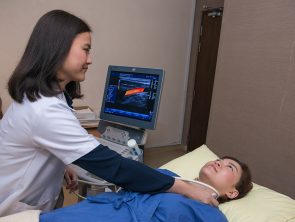
Carotid Intima-Media Thickness (Carotid IMT) Ultrasound
Measurement of carotid IMT in the carotid artery with ultrasound is a non-invasive, sensitive, and reproducible technique for identifying and quantifying subclinical vascular disease and for evaluating Cardiovascular Disease (CVD) risk – heart attacks and strokes and also to predict future cardiovascular disease (heart attacks and strokes) risk.
Who should get a Carotid IMT Ultrasound Test?
- Patients at intermediate CVD risk (i.e. 6%-20% 10-year risk for myocardial infarction or coronary heart disease death who do not have established coronary heart disease or coronary disease risk equivalent conditions)
- Family history of premature coronary heart disease (heart attacks and strokes) in a first-degree relative;
- Younger than 60 years old with severe abnormalities in a single risk factor who otherwise would not be candidates for pharmacotherapy;
- Women younger than 60 years old with at least two coronary heart disease risk factors.
- If the level of aggressiveness of therapy is uncertain and additional information about the burden of subclinical vascular disease or future coronary heart disease (heart attacks and strokes) risk is needed.
- Reclassify patients at intermediate risk, discriminate between patients with and without prevalent coronary heart disease, and predict major adverse coronary heart disease (heart attacks and strokes) events.
Coronary Heart Disease (CVD) risk factors (Risks for developing heart attacks and strokes):
- Hypertension
- Diabetes
- Raised total cholesterol, raised triglycerides, raised LDL cholesterol, raised ratio total cholesterol/HDL and reduced HDL cholesterol
- Overweight and obesity
- Smoking
- Males above 35 years old and post-menopausal women.
- Family history of heart attacks and stroke.
- Medical history of arrhythmias (irregular heartbeat)
- Transient Ischemic Attack (TIA)- minor stroke
- Peripheral Arterial Disease (PAD) – narrowing or blockages of arteries in the legs.
When does atherosclerosis starts?
The atherosclerotic vascular disease begins in childhood and progresses over decades. Symptomatic, clinical cardiovascular disease (CVD) events generally occur when atherosclerosis progresses to flow-limiting disease that causes ischemia, or when a thrombus forms on an existing plaque as a result of rupture or erosion.
Although not everyone with underlying atherosclerotic plaque will experience a clinical CVD event, the greater the degree of subclinical atherosclerosis, the greater the risk for future cardiovascular events.
To prevent death and morbidity from CVD, there is great interest in identifying asymptomatic patients at high risk who would be candidates for more intensive, evidence-based medical interventions that reduce CVD risk.
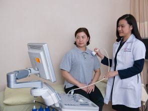
Echocardiography
This is a common non-invasive investigation tool for heart conditions. It applies ultrasound waves to generate images of the structures of the heart. It allows real-time visualization of the heart and the large blood vessels connected to it.
With the use of Doppler ultrasound technology, we are also able to interrogate the function of heart valves, derive blood pressures of different heart chambers and assess the functions of the heart.
How do I prepare for an Echocardiography examination?
There is no special preparation needed for this painless, non-invasive procedure. Clothing from the upper body is removed and covered by a gown or sheet to keep you comfortable and maintain the privacy of females. The patient then lies on an examination table.
How is an Echocardiography examination done?
ECG electrodes are attached to the body to allow the timing of the heart events to the ECG cycle. A transducer (a probe that transmits and receives ultrasonic waves) is placed over the chest or region of interest. A colourless gel is applied to the probe before scanning to improve the image quality.
The echo technologist or cardiologist would acquire images from different parts of the chest to obtain views from different angulations. You may be advised to assume certain body positions for better imaging of the different heart or non-heart structures.
How long does an Echocardiography examination take?
An evaluation of a normal heart generally takes about 10-15 minutes. However, a more detailed interrogation of abnormal findings may take a bit longer.
What information can I obtain from an Echocardiography examination?
- Size of the heart chambers and thickness of heart muscles
- The function of the heart
- Heart valve structures and their functions
- Blood pressure in the heart and lungs
- Extra-cardiac structures eg. Pericardium (outer lining of the heart), aorta (a large blood vessel that arises from the heart)
- Presence of abnormal heart structures e.g. Hole-in-the-heart, growths involving the heart
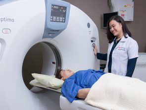
(Heart CT Scan) Calcium Score
Calcium is a marker of coronary artery disease, the amount of calcium detected on a heart CT scan is a helpful diagnostic tool. The findings of heart CT are expressed as a calcium score. Another name for this test is Coronary Artery Calcium Scoring.
CT Scan is a noninvasive, painless medical test that helps doctors diagnose and treat medical conditions. A heart CT Scan is a non-invasive way of obtaining information about the location and extent of calcified plaque in the coronary arteries – the vessels that supply oxygen-containing blood to the heart wall.
Plaque is a build-up of fat and other substances, including calcium, which can, over time, narrow the arteries or even close off blood flow to the heart. The result may be painful angina in the chest or a heart attack.
How should I prepare for the procedure?
No special preparation is necessary in advance of a heart CT scan. You may continue to take your usual medications but should avoid caffeine and smoking for 4 hours before the exam. If your heart rate is 90 beats a minute or higher, you may be given a drug to slow the rate in order to obtain accurate CT images.
You should wear comfortable, loose-fitting clothing to your exam. You may be given a gown to wear during the procedure. Metal objects including jewellery, eyeglasses, dentures and hairpins may affect the CT images and should be left at home or removed prior to your exam. You may also be asked to remove hearing aids and removable dental work.
Women should always inform the doctor or technologist if there is any possibility that they are pregnant.
Non-invasive CT Coronary Angiography
CT Angiography is a safe outpatient procedure that uses specially designed x-rays and intravenous contrast to see the detailed anatomy of the blood vessels throughout the body. It is most frequently utilized in the evaluation of arteries in the head, neck, chest, abdomen and legs. Coronary CT angiography refers to the scanning procedure of the arteries of the heart i.e. those involved in ischemic heart disease like heart attacks.
How safe is a CT Coronary Angiography scan?
There are certainly radiation issues with this modality. The effective dose is almost similar to that of an invasive heart catheterization (5-8 mSv). Contrast use would entail the risk of allergic reaction in a small proportion of the population. Therefore, not all persons would benefit from this test. Please discuss with the doctor the proper indications and the risk-benefit analysis of performing such a scan.
Who should have a CT Coronary Angiography scan?
Coronary CT Angiography is ordered by the doctor if there is a need to establish the presence of significant coronary artery disease. It may be indicated if initial heart screening tests e.g. Stress ECG tests or nuclear heart scans are abnormal. It is also used for the follow-up of patients after coronary angioplasty and stenting, post coronary bypass grafting surgery.
How is the CT Coronary Angiography done?
Intravenous access needs to be obtained. You will be asked to lie down on the CT scanning table. Breath-holding instructions will be given. Often the whole heart can be imaged within 5 heartbeats. Contrast injection is necessary and sometimes, you may need medication to slow down the heart rate.
Following image acquisition, our radiographer and doctor will process and report on the images. The results will be discussed with you.
How can I prepare for a CT Coronary Angiography scan?
You need only to fast for about 4 hours before the scan. Please refrain from caffeinated drinks e.g. coffee or tea on the day of the examination.
How long does a CT Coronary Angiography Scan take?
Although the actual heart scanning takes only 5-8 seconds, the whole scanning procedure may take about 15-20 minutes from preparation to completion. You would be able to leave immediately after the procedure and would be able to resume normal activities.
How good is a CT Coronary Angiography?
The most advanced multi-detector CT today is a 128-detector row scanner. By using very fast gantry rotation speed and large volume detector coverage, we are able to obtain detailed visualization of the coronary tree. This is a very promising new technology. The latest data are reporting excellent results.
A recent study, presented in March 2005 at the annual meeting of the American College of Cardiology, compared the capacity of 128-slice heart CT versus coronary angiography to detect significant coronary artery stenosis (defined as more than 50 per cent lumen diameter reduction). 30 patients with stable angina or acute coronary syndrome were enrolled in the study; individuals unable to hold their breath for less than 15 seconds were excluded from the study. The heart was scanned following intravenous injection of contrast material and analyzed by two observers who were not aware of the results obtained from invasive angiography.
Most patients received a beta blocker before the study, and all coronary arteries were compared between coronary CT and selective angiography. Compared with selective angiography, 128-channel heart CT showed a sensitivity of 96% and specificity of 89% when detecting significant stenosis (narrowing). The authors concluded that 128-channel heart CT could reliably detect significant coronary stenosis in patients with stable angina or acute coronary syndrome (Source: Mollet, et. al., presentation #1054-83, ACC ‘05).
A more recent analysis (Hoffman et al., JAMA 2005; 293:2471-2478) showed an MDCT sensitivity of 95% and specificity of 98%.
Is CT Coronary Angiography an alternative to Catheter Angiography?
Heart catheterization remains the gold standard for diagnosing coronary artery disease. However, the setback with this approach lies in the fact that it is invasive, requires a longer hospital stay and has complications, e.g., bleeding, and pain and it costs more.
Compared to Catheter Angiography, CT Angiography is a less invasive and more patient-friendly procedure. Although contrast is injected through an artery in Catheter Angiography, CT Angiography contrast is injected into a vein which is technically less difficult and has a very low risk of complication. As a result, the patients typically leave immediately following the procedure and can resume normal activities.
Heart CT also offers the advantage of being able to visualize atherosclerotic plaque composition. Heart catheterization only opacifies the arterial lumen.
What does it mean if my scan is positive or equivocal?
Your scan would be able to tell if there is an atherosclerotic narrowing of the coronary arteries. It can also estimate the degree of obstruction. Based on these findings, the cardiologist will advise you further on the subsequent management.
Sometimes, the presence of excessive calcium or certain stents in the arteries may interfere with the proper interpretation of the scans and hence reduce their accuracy.
Does a negative scan mean that I am free from Coronary Arterial Disease?
Having a normal scan is an excellent result. The negative predictive value of a 128-slice MDCT Coronary Angiography is about 99-100%. This means that if the scan result is normal, the likelihood of this being true is nearly 100%.


![Gastroscopy (OGDS) [Consultation by Gastroenterologist]](https://lifecarediagnostic.com/wp-content/uploads/1970/01/Gastroscopy-Product-Thumbnail-ENGLISH-FA-300x186.jpg)